

Medical imaging
OpenReadout reads CT, MR, PET, ultrasound, X-ray and nuclear medicine images from DICOM files and folders, NIfTI and Analyze volumes from neuroimaging, and Bruker ParaVision experiments from preclinical MRI. The crate is openreadout-medical. Format notes: DICOM, NIfTI and Analyze, Bruker ParaVision.
What you get
- One image per series. Point
infoat a DICOM file, a series or study folder, or a DICOMDIR. Slices are grouped by series and sorted along the slice normal into a volume (z), and repeated volumes or cine frames become t. Enhanced multi-frame files, segmentations (one channel per segment) and whole-slide microscopy (a pyramid, read tile by tile) are read too. - Real units. Values come out rescaled: CT in Hounsfield units, PET and parametric maps in their stored units (
extra.value_unit,extra.rescale). Voxel sizes are inphysical_size, andextra.orientationsays which way columns, rows and slices run (L/R, A/P, S/I) and which plane the slices are in. - Personal data stays out. Patient names, IDs, birth dates and other identifying DICOM elements and ParaVision subject parameters are shown as
withheld, ininfoand in--view full.extra.personal_datalists which elements hold them, andindexflags these files in a lab share. - Compressed DICOM. Implicit and explicit VR, big endian, deflate, RLE, JPEG (baseline, extended, lossless), JPEG-LS and JPEG 2000 (including HTJ2K), all decoded in pure Rust.
Pictures
preview shows a slice the way a radiologist expects: the plane the scan was acquired in, patient left on the right of the picture, the file’s display window (or the soft-tissue window for CT), and a scale bar in mm. --contrast window:bone (or brain, lung, soft-tissue, …) picks a preset, and --view coronal, sagittal or orthogonal cuts the volume along another plane.
openreadout preview ct-series/ --view orthogonal -o ct.jpg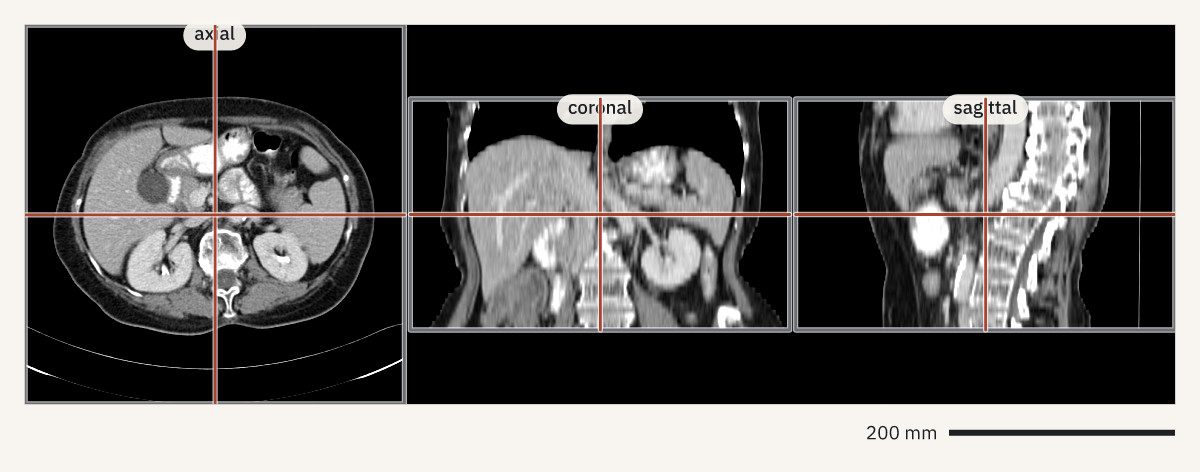
Contrast-enhanced abdominal CT, 41 slices of 5 mm (StageII-Colorectal-CT collection, The Cancer Imaging Archive, CC BY 4.0).
A NIfTI volume is shown the same way. Voxels that are not square are stretched to their true shape.
openreadout preview sub-01_T1w.nii.gz --view sagittal -o t1.jpg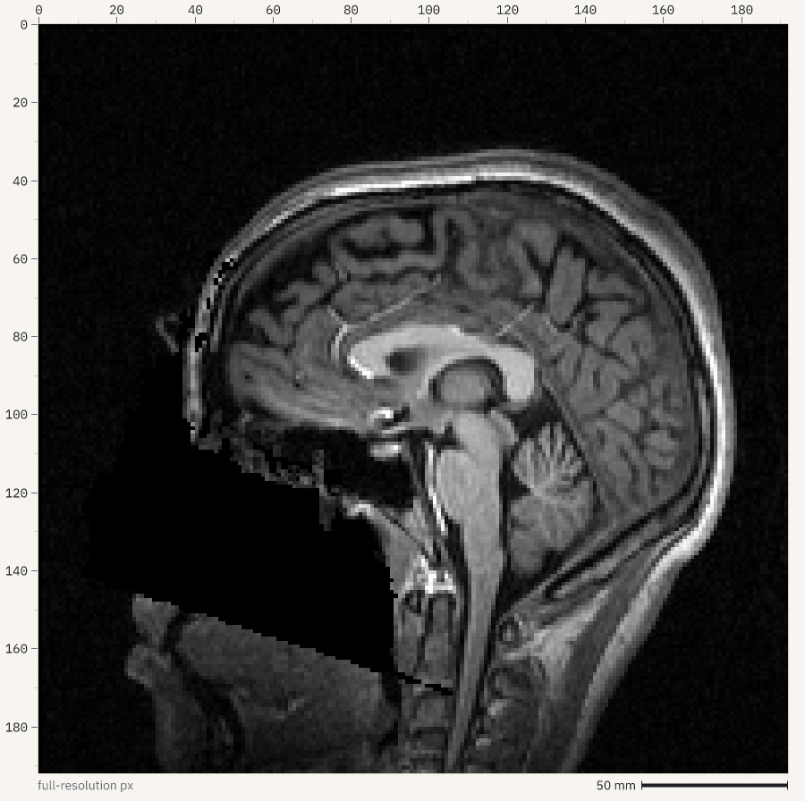
T1-weighted MRI, OpenNeuro ds000001 (CC0).
Numbers
stats reports values in the image’s units, so a region of a CT gives Hounsfield units. A plot series with show: profile draws the values along a line:
openreadout plot '{"data": {"file": "ct-series/", "show": "profile", "line": [[0, 256], [512, 256]]}}' -o profile.png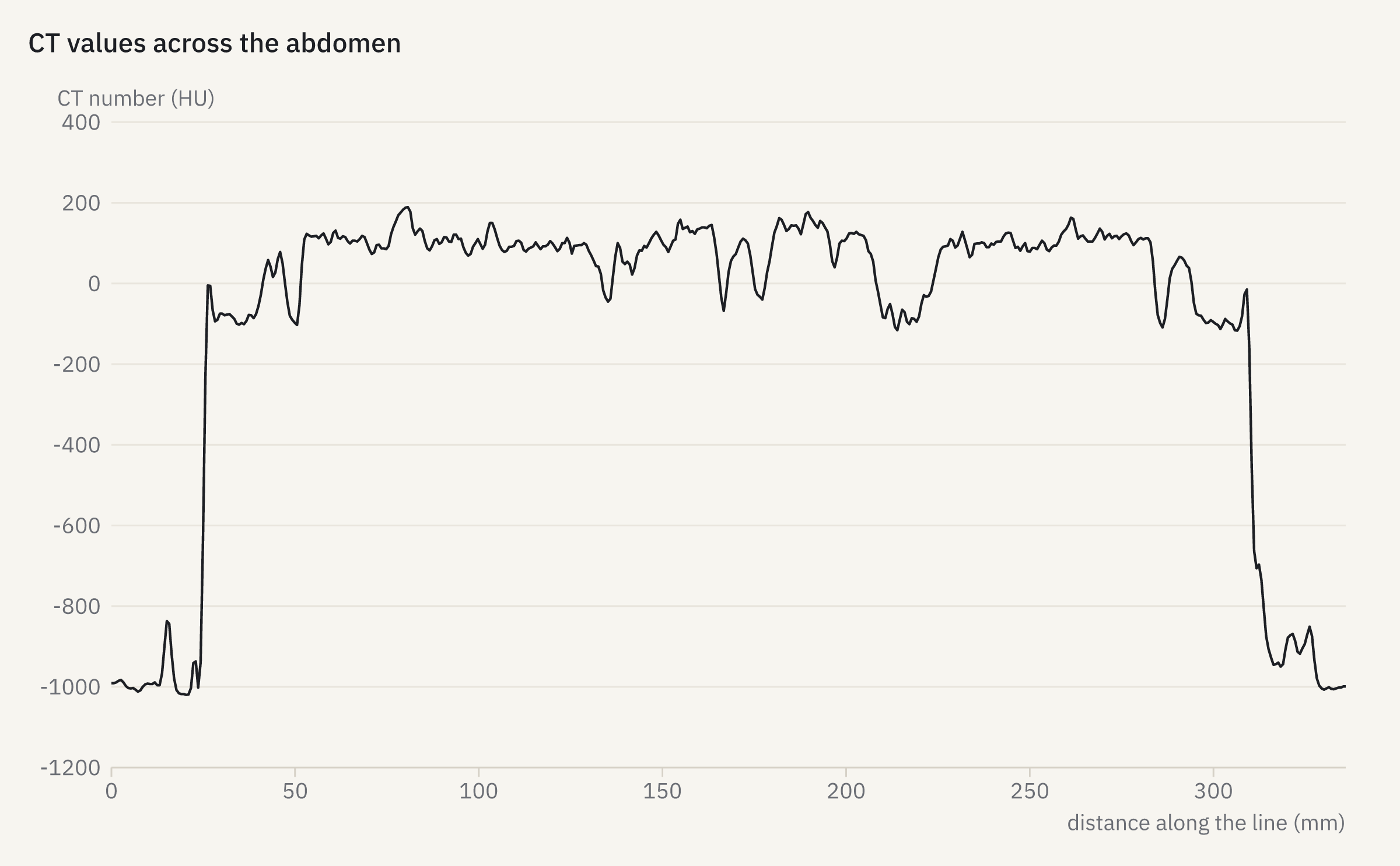
Export
export -o scan.nii.gz writes NIfTI-1 with the voxel size and the scanner orientation (from the DICOM geometry or the source NIfTI header). OME-TIFF and OME-Zarr work as for any other image.
Not read yet
MINC, NRRD and FreeSurfer MGH volumes are candidates for later. Raw k-space files (Siemens .dat, Philips .raw, GE P-files) and ECAT are out of scope.